
Nicholas George Zaorsky, Ninad Patil,1 Gary Mitchel Freedman,2 and Madalina Tuluc3.
- Department of Pathology, Fox Chase Cancer Center, Philadelphia, USA.
- Department of Radiation Oncology, University of Pennsylvania, Philadelphia, USA.
- Department of Pathology, Jefferson Medical College, Thomas Jefferson University, Philadelphia, USA.
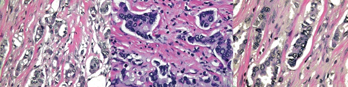
Abstract: On a pathological specimen of breast cancer cells, retraction artifact during histological processing mimics true lymphovascular invasion (LVI). The accurate determination of the presence or absence of LVI is a factor in determining risk of having a positive sentinel node, or having additional positive axillary nodes after a positive sentinel node biopsy in women with early-stage breast cancer. The determination of nodal risk influences the decision of the treating physicians as to whether a sentinel node biopsy or completion axillary dissection is necessary. On slide preparation, ideal factors favoring true LVI include: a definite endothelial lining, with endothelial nuclei that seem to protrude into the lymphatic space; invasion in one lymphatic vessel (LV) lumen with nearby cancer glands that have minimal or no retraction; a tumor embolus in a LV clear lumen with outside nearby tumor bulk; a tumor embolus that is different in shape than its surrounding clear LV space; and a positive stain for fibrin, CD31, or CD34 on tumor embolus periphery.
Keywords: Breast neoplasms; Diagnosis; Pathology.
J Breast Cancer. 2012 Dec;15(4):478-480. English.
Published online Dec 31, 2012. Artículo completo de libre descarga en la página web.
© 2012 Korean Breast Cancer Society. All rights reserved.
